Intraoral Scanning vs. Analog Impressions: The Digital Shift in Modern Dentistry
In dental practice, precision is paramount. Whether for prosthodontics, orthodontics, or implantology, the accuracy of impressions directly impacts treatment outcomes. Traditionally, analog impressions have been the gold standard, but intraoral scanning (IOS) is rapidly redefining clinical workflows. Understanding the nuances of each method is essential for clinicians looking to optimise accuracy, efficiency, and patient experience.
Analog Impressions: A Time-Tested Approach
Analog impressions involve using materials like polyvinyl siloxane (PVS) or polyether to create a physical mould of the dentition. While time-tested, the workflow presents several considerations:
- Clinically reliable for full-arch cases and edentulous spans.
- Material familiarity and lab compatibility make it a universally accepted technique.
- Technique sensitivity can compromise results — errors in mixing, tray seating, or removal are common.
- Patient discomfort is a major drawback — bulky trays and viscous materials often trigger gag reflexes.
- Susceptible to dimensional instability due to temperature changes, humidity, or delayed pouring.
- Multiple manual steps (pouring, trimming, scanning) increase chairside time and risk of inaccuracies.
- Prone to volumetric changes due to polymerisation shrinkage (0.05–0.3%) and thermal expansion.
- Errors may arise from hydrophilicity, tray selection, or impression material handling.
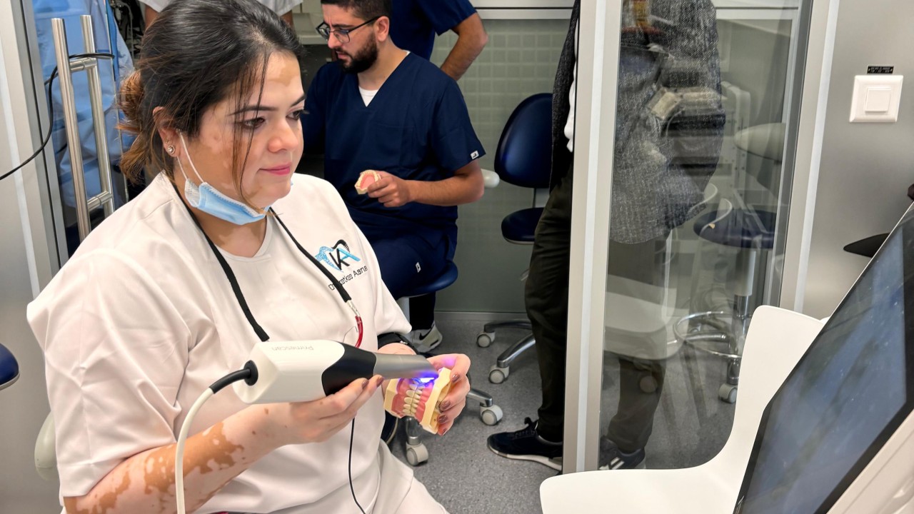
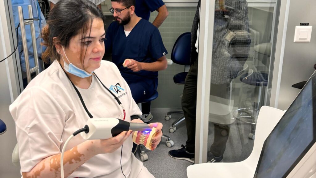
Intraoral Scanning: The Digital Evolution
Intraoral scanners (IOS) use structured light or laser technology to digitally capture the dental anatomy in 3D. The shift towards IOS is driven by:
- High-resolution accuracy with reduced operator-dependent variability.
- Immediate visual feedback, enabling real-time error correction.
- Vastly improved patient comfort — no impression trays, no gag reflex.
- Seamless integration with CAD/CAM systems for same-day restorations and digital treatment planning.
- Instant data transfer to labs — no shipping delays, no need for physical models.
- Efficient record keeping — digital files are easy to store, replicate, and compare over time.
- Supports a sustainable practice by eliminating disposable materials like trays, PVS, and plaster.
- Eliminates errors from material shrinkage, expansion, or improper disinfection.
- Offers real-time tissue evaluation, preventing compression artefacts seen with PVS.
- Trueness reported within 10–30 μm, often outperforming PVS in controlled environments.
- CAD/CAM-milled restorations from digital impressions show marginal gaps between 25–50 μm, within clinically acceptable thresholds (<120 μm).
- Enables more precise digital occlusion assessment (dynamic and static), surpassing wax and bite paste articulation.
- Further evaluation of occlusion using digital software is more predictable than traditional articulating paper checks.
That said, IOS has its considerations:
- Initial investment in hardware, software, and training can be substantial.
- Requires operator training and familiarity with digital workflows.
- Full-arch scans, particularly in edentulous or mobile soft tissue cases, require precise technique and appropriate scanner selection.
- Occlusal accuracy must be carefully managed — bite registration protocols must be strictly followed.
- Cross-arch distortion can be a concern in complex full-arch rehabilitations — case selection remains key.
Scientific Insights and Clinical Evidence
- Comparative studies suggest IOS offers comparable or superior accuracy for single-unit and short-span FDPs.
- A systematic review (Mühlemann et al., 2019) found IOS to demonstrate superior reproducibility and marginal fit in single and short-span restorations, while PVS was marginally more reliable for full-arch cases.
- Meta-analyses (Mangano et al., 2020) show IOS exhibits lower linear distortion compared to PVS for implant-supported restorations.
- Hybrid workflows combining digital impressions and conventional elements can achieve better passive fit of prostheses in select cases.
While analog impressions will continue to hold relevance in certain clinical contexts, the future is undeniably digital. As intraoral scanning becomes more accessible — with AI-enhanced software, faster scan speeds, and affordable options — adoption is no longer a question of “if” but “when.”
For practitioners starting out, a phased integration—beginning with single-unit crowns or diagnostic models—can ease the transition. Over time, expanding into implant and full-arch applications will come naturally as proficiency grows.
As AI-powered IOS, diagnostic assistance, and dynamic occlusion tracking continue to evolve, digital impressions will soon surpass conventional methods in accuracy, efficiency, and long-term predictability. It’s not far from adoption at every clinic. However, case selection remains key, particularly in full-arch rehabilitations where cross-arch distortion is still a concern.
And what’s my verdict on this? Adopting intraoral scanning is more than a technological shift — it is a commitment to clinical excellence, efficiency, and patient-centric care. The future of digital dentistry lies in AI-enhanced scanning, INTRAORAL PHOTOGRAMMETRY, and real-time dynamic occlusion tracking, ensuring a fully digital workflow from planning to prosthesis delivery. In a profession where precision defines success, going digital is not just an upgrade — it’s a strategic move to future-proof your practice.